Login
LoginElectroencephalogram (EEG)

- What is an EEG?
- What does an EEG detect?
- When is an EEG used?
- How to prepare for an EEG
- How an EEG is done
- Risks of an EEG
- Usefulness of EEGs
What is an EEG?
EEG stands for electroencephalogram. In its commonest form, it is a measurement of the electrical activity of the brain measured from an array of electrodes on the scalp. There are other forms of EEG that are more invasive methods of recording but these are not widely used. By recording the electrical signals, the resulting traces are known as the EEG. The EEG is one of the commonest investigations for brain pathology. Over the years, the clinical utility of this test has declined because of the development of sensitive neuroimaging techniques, but it still fulfils an important role.
 trace") |
| An EEG trace. |
What does an EEG detect?
The waveforms in the EEG are generated by impulses near the communication between neurons or nerves in the brain. They are picked up by the electrodes on the surface of the scalp, and therefore the standard test may not be able to show smaller activity that occurs deep within the brain.
When is an EEG used?
An EEG is done for the following reasons:
- As an aid to the diagnosis of epilepsy: This is the most useful and important test in confirming a diagnosis of epilepsy. However a normal EEG does not exclude the diagnosis of epilepsy. The test can help classify the type of epilepsy and also differentiate it from non-epileptic events.
- The assessment of impaired consciousness or altered mental state
- To evaluate cognitive impairment and dementia.
- Sleep disorders such as narcolepsy.
- To monitor brain activity when a person is under general anaesthesia.
- Much less frequently nowadays, to localise suspected lesions of the brain such as tumour, inflammation, infection, or vascular events like stroke. It remains an important adjunct in the detection of temporal lobe involvement in encephalitis, for example due to herpes infection.
How to prepare for an EEG
") Before the test, a complete medical history will be taken by the doctor ordering the test to determine if the EEG is really necessary and indicated. After that, the doctor or laboratory will advise the patients of the following:
Before the test, a complete medical history will be taken by the doctor ordering the test to determine if the EEG is really necessary and indicated. After that, the doctor or laboratory will advise the patients of the following:
- Before the day of EEG, medications that can affect brain activity may be ceased, but this is not usually necessary. These can include sedatives, tranquilisers, sleeping tablets and medications for epilepsy. These can affect the test but it is very important that you consult your doctor before you stop them.
- At least 8 hours before the test, foods that contain caffeine (coffee, tea, cola, chocolate) should be avoided.
- Hair styling products (hairspray or gel) should be avoided during the day of test as this impedes electrode placement on the scalp.
- Shortly before the procedure, a small meal should be eaten to maintain blood glucose level to the brain.
- If a sleep-deprived or sleep EEG is to be obtained, the patient might be asked not to sleep for some hours or for all of the night before the test.
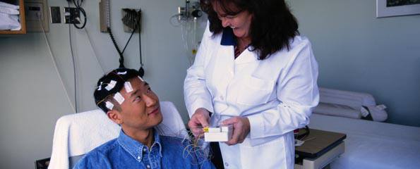
How an EEG is done
The standard EEG takes about 1 hour. The patient will be positioned on a bed or chair. After that, 16-20 electrodes will be attached to the scalp by using gel. The technician/nurse/doctor might ask the patient to breathe deeply for many times in a short period to provoke or enhance any abnormalities on the EEG. Additionally, there may also be a series of flashes given with a light strobe. If a sleep EEG is performed, it may last for 2-3 hours. If an ambulatory EEG is done, it typically lasts for 24 hours, but the patient is allowed to perform daily activities. After the test, the patient is usually allowed to go home without the need for a recovery period provided the test went smoothly. If the patient has a high possibility of having a seizure, someone should take care of the patient. The results will be discussed with the patient by the doctor or referring clinician.
") |
| (Image courtesy of Dr K Ng) |
Risks of EEGs
An EEG is a non-invasive procedure, hence very few risks are associated with it. Occasionally and sometimes because a patient may be withheld from antiepileptic medications or had medications reduced, the patient may experience clinical seizures during the procedure. However, most patients will not be told to stop their medication for seizures. To provoke abnormalities in the EEG, the patient may be asked to do certain activities such as deep breathing. While not particularly uncomfortable (sometimes accompanied by dizziness), an abnormality on the EEG is what is desired during the procedure. It must be remembered that EEG does not provide stimulation to the brain – it merely detects and receives electrical signals from the brain.
Usefulness of EEGs
This test is usually totally non-invasive and has very few side effects apart from potential seizure activities during the procedure. Hence the doctor will almost always recommend this test when it is needed.
Article kindly reviewed by:
Associate Professor Karl Ng MB BS (Hons I) FRCP FRACP PhD CCT Clinical Neurophysiology (UK) Consultant Neurologist – Sydney North Neurology and Neurophysiology (download referral form and map); Conjoint Associate Professor – Sydney Medical School, University of Sydney; and Editorial Advisory Board Member of the Virtual Neuro Centre.
References
- Vrocher D, Lowell MJ, Plantz SH. Electroencephalography (EEG) [online]. eMedicineHealth; 2005 [cited 22 October 2005]. Available from: [URL link]
- Bradley WG, Daroff RB, Marsden CD, et al (eds). Neurology in Clinical Practice (3rd edition). Philadelphia: Butterworth-Heinemann; 2000.
- Schachter SC. Evaluation of the first seizure in adults [online]. UpToDate; 2005 [cited 22 October 2005]. Available from: [URL link]
- Deuschl G, Eisen A (eds). Recommendations for the Practice of Clinical Neurophysiology: Guidelines of the International Federation of Clinical Neurophysiology. Electroencephalogr Clin Neurophysiol Suppl. 1999;52:1-304. [Abstract]
Dates
Tags
Created by: